Figure 1.

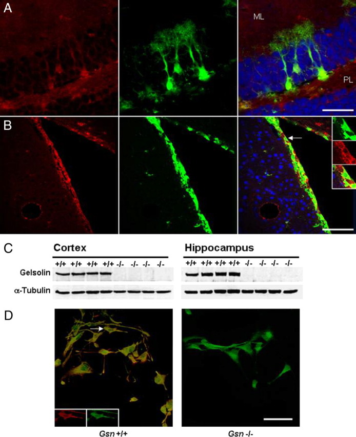
Gelsolin expression in adult neural progenitor cells. Characterization of gelsolin expression (red) in the hippocampal dentate gyrus (A) and in the subventricular zone (B) of 3- to 4-month-old nestin-GFP (green) reporter mice. Blue, Neuronal marker NeuN. A, Confocal image demonstrating gelsolin expression in the cell bodies and processes of radial glia-like nestin-GFP cells in the hippocampal dentate gyrus. Note that, in the granule cell layer, gelsolin is also expressed in neurons. Furthermore, there is widespread gelsolin staining in the neuropil of the molecular layer (ML) and of the polymorphic layer (PL). B, Gelsolin expression in neural progenitors of the subventricular zone. The insets show higher magnification of the nestin-GFP cell marked by arrow. C, Western blot analysis of protein extracts with gelsolin antibody confirms lack of gelsolin in Gsn−/− mice. D, Whereas cultures of neural progenitors derived from Gsn+/+ mice show gelsolin immunoreactivity, neural progenitors from Gsn−/− mice lack gelsolin. Green, Nestin protein. The insets show cell marked by arrow in single channels with separate wavelengths. Scale bars: A, 50 μm; B, 100 μm; D, 50 μm.
