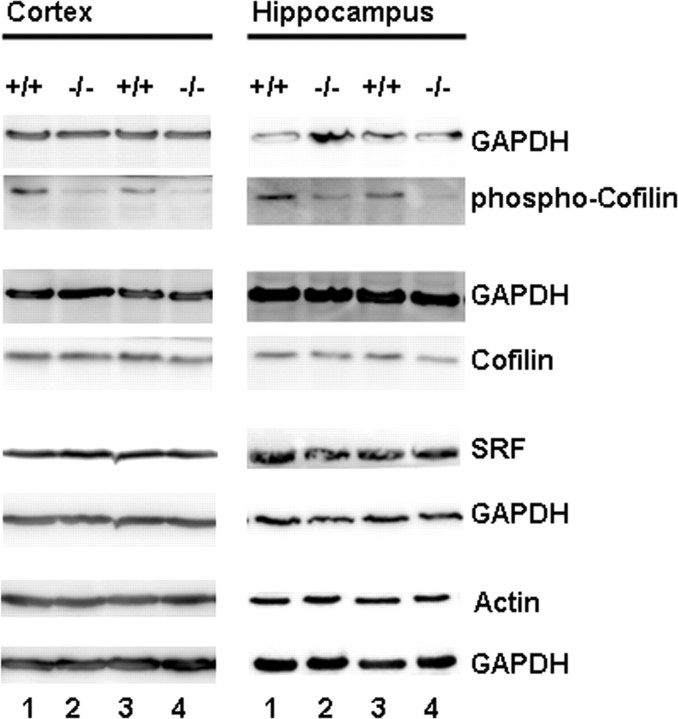
Figure 9.
Dephosphorylation of cofilin in Gsn−/− mice. Western blot of protein extracts from hippocampus and frontal cortex of control (+/+) and gelsolin-deficient animals (−/−) probed with antibodies for actin, cofilin, phospho-cofilin, and SRF. Comparable loading of protein is confirmed by GAPDH staining.

