
Figure 2.
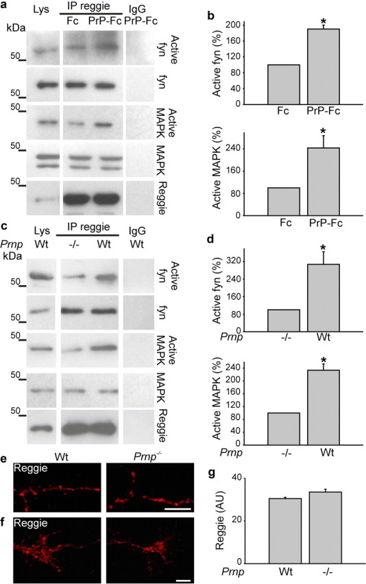
PrP regulates reggie-associated fyn and MAPK activity. a, HPL3-4 cells transfected with PrP were incubated live with Fc or PrP-Fc. Reggie immunoprecipitates from cell lysates (Lys) were probed by Western blot with Abs against total and active forms, respectively, of fyn and MAPK. Nonimmune rabbit IgG was used as control. Western blots for reggie were included as loading control. b, Activated fyn and MAPK were increased in reggie immunoprecipitates when stimulated with PrP-Fc. c, Reggie immunoprecipitates from 0 to 3 d wt and PrP−/− mice were probed with Abs against total and active fyn and MAPK, respectively. Immunoprecipitation with nonspecific IgG was used as control. Active fyn and MAPK were reduced in PrP−/− brains. Histograms (b, d) show quantification of the blots with values for PrP−/− and Fc stimulation, respectively, set to 100%. Mean values ± SEM (n > 6) are shown. *Statistical significance, p ≤ 0.05, paired Student's t test. e, f, PrP−/− and wt hippocampal neurons, when labeled with reggie Ab, show no apparent difference in the distribution of reggie in PrP−/− and wt neurons. Scale bar, 5 μm. g, The histogram corresponding to e and f shows mean intensity of reggie. Mean values ± SEM (n > 50) are shown. *Statistical significance, p ≤ 0.05, paired Student's t test.