
Figure 3.
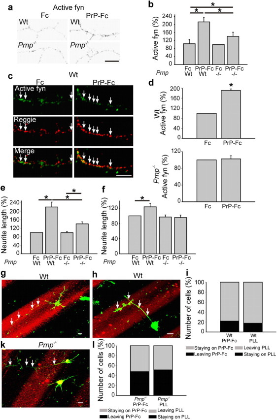
PrP-Fc leads to an increase in neurite length, reggie-associated fyn activation, and preference of neurite extension. a, Wt and PrP−/− hippocampal neurons were incubated live with Fc or PrP-Fc, fixed, and labeled with Abs against active fyn. Immunofluorescence signals are shown in black-and-white to ease recognition of the difference in immunolabeling intensities between groups. Scale bar, 10 μm. b, Application of PrP-Fc increased the level of active fyn in wt and in PrP−/− neurons as indicated in the histogram. Mean values ± SEM (n > 100 neurons). *Statistical significance, p ≤ 0.05, one-way ANOVA test. c, WT and PrP−/− hippocampal neurons were incubated live with Fc or PrP-Fc to cluster PrP. Cells were fixed and labeled with active fyn and reggie Abs. The amount of active fyn in reggie clusters was reduced in PrP−/− neurons. Scale bar, 10 μm. d, The histogram shows mean (±SEM; n ≥ 50 neurons) intensity of active fyn in reggie clusters. *Statistical significance, p ≤ 0.05, paired Student's t test. e, f, PrP−/− and wt hippocampal neurons growing on poly-l-lysine (e) and laminin (f) were incubated with PrP-Fc or Fc as control and lengths of the longest neurites were measured. e, On poly-l-lysine, wt neurons stimulated with PrP-Fc show a 120% increase in neurite length over Fc-treated neurons, and a 64% increase compared with PrP-Fc-treated PrP−/− neurons. f, On laminin, PrP-Fc increased neurite length of wt neurons by 24% over Fc, but had no effect on neurite length in PrP−/− neurons. Mean values ± SEM are shown (n > 100). *Statistical significance, p ≤ 0.05, one-way ANOVA test. g, h, When seeded on a substrate consisting of alternating lanes of poly-l-lysine and PrP-Fc (red), wt neurons showed a preference to reside and to extend neurites (arrows) on the PrP-Fc stripe, whereas neurites (arrows) of PrP−/− neurons (k) did not exhibit a preference for either substrate and crossed the stripes. Scale bar, 10 μm. i, The histograms reflect the number of wt neurons that resided on PrP-Fc and either kept all processes on PrP-Fc (staying on PrP-Fc) or sent processes to poly-l-lysine (leaving PrP-Fc) as opposed to wt neurons that resided on poly-l-lysine and either kept their neurites on poly-l-lysine (staying on PLL) or sent processes to the PrP-Fc lane (leaving PLL). These values are compared with the behavior of PrP−/− neurons (l) as is reflected by the corresponding histogram.