
Figure 7.
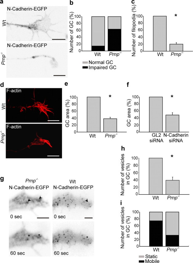
PrP−/− hippocampal neurons exhibit abnormal growth cones. a–h, Wt and PrP−/− neurons were transfected with N-cadherin-EGFP or stained with phalloidin, to visualize the cell and growth cone morphology. Growth cones of PrP−/− neurons were abnormally small (a, b, d, e), had significantly fewer filopodia (c), and were poor in N-cadherin cargo vesicles (h). e, f, Growth cone size was significantly reduced in PrP−/− neurons much as in siRNA N-cadherin-treated neurons. When monitored by time-lapse recordings (g, i), the N-cadherin vesicles were by far less motile in PrP−/− neurons than in their wt counterparts (i). Scale bar, 5 μm. Mean values ± SEM (n > 100) are shown. *Statistical significance, p ≤ 0.05, paired Student's t test.