
Figure 5.
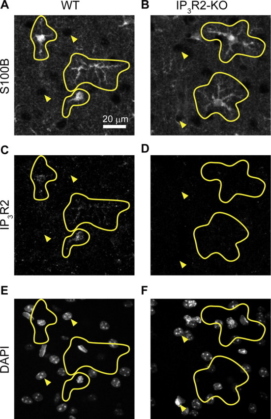
Double immunohistochemistry for S100B (astrocyte) and IP3R2 with DAPI staining in the barrel cortex. A, B, Immunohistochemistry for the astrocytic protein S100B in WT and IP3R2-KO mouse barrel cortex. C, D, Immunohistochemistry for IP3R2 of the same sections as A and B, respectively. E, F, DAPI counterstaining of the same sections as A and B, respectively. Astrocytic morphology was delineated in yellow based on S100B immunohistochemistry. Filled triangles point to nonastrocytic (presumably neuronal) cells. Immunoreactivity against IP3R2 is predominantly localized in astrocytes.