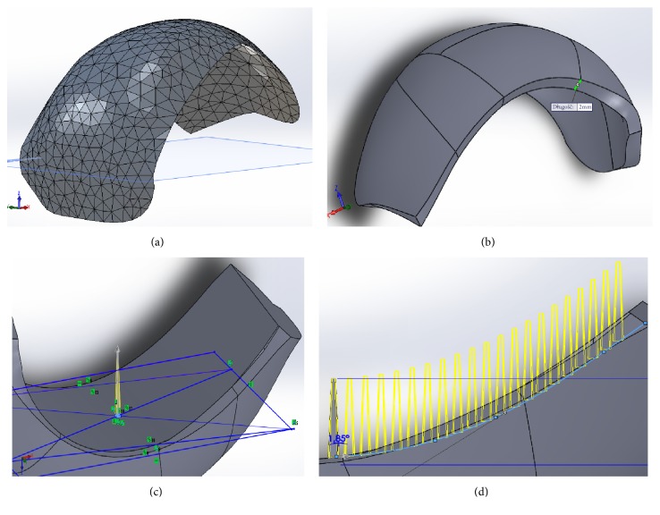
Figure 3.
The main stages of CAD-modeling of working prototype of partial RKA endoprosthesis: (a) 3D triangular representation of the articular surface of lateral condyle of the femur as imported to the CAD software; (b) view after adding thickness in the normal direction inwards; (c) a screenshot showing the manner of determining of the initial spike of the MSC-Scaffold; (d) a screenshot showing a preview of the multiplying of spikes using the “curve driven pattern” tool.

