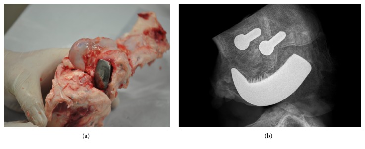
Figure 9.
The retrieved specimen vs. its X-ray radiogram: (a) the exemplary specimen of operated swine knee joint with the implanted working prototype of partial knee resurfacing endoprosthesis harvested at 8 weeks after implantation; (b) the exemplary 2D digital X-ray radiogram of the resected at 8 weeks after implantation swine knee joint showing spaces between the spikes of the MSC-Scaffold of the implanted RA endoprosthesis working prototype penetrated with bone tissue and the two trabecular bone screws used for the reattachment of the femoral part of the lateral collateral ligament of the swine knee joint.

