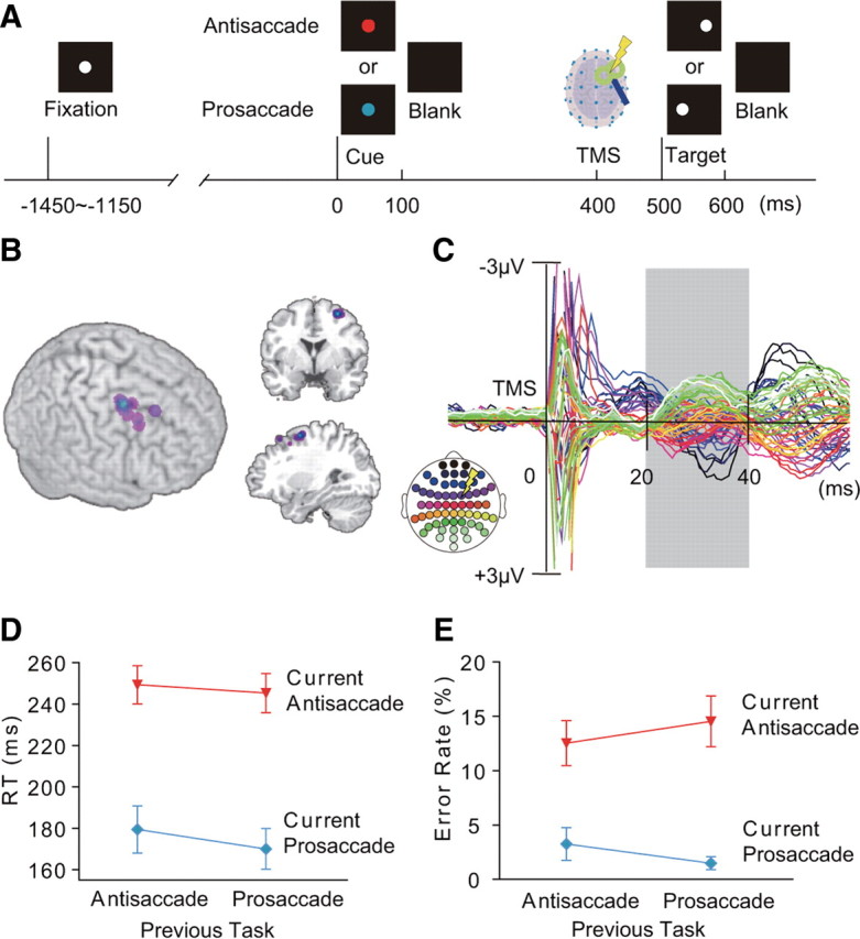
Figure 1.

Cued saccade task and TMS-EPs. A, Time line of the task. Subjects made prosaccade or antisaccade according to a pretarget cue (red cue: antisaccade, cyan cue: prosaccade). B, Position of the FEF-TMS rendered on 3D surface of the MNI template brain (left), coronal section at y = 2 (upper right), and sagittal section at x = 30 (lower right). C, TMS-EPs averaged across all conditions and all subjects, displayed from −20 to 60 ms of TMS. TMS-EPs were calculated by subtracting the EEG waveforms on no-TMS trials from those on TMS trials. The analysis was focused on the time window of 20–40 ms after TMS (gray shading). Colors of the waveforms correspond to the electrode positions on the scalp (inset at lower left). A yellow symbol of lightening bolt in the inset indicates scalp position at which TMS was applied. D, E, Latency of saccade onset (RTs) (D) and error rates (E), separately shown for the four conditions based on the task on the previous and current trials. Mean and SE across subjects are shown.
