Figure 5.
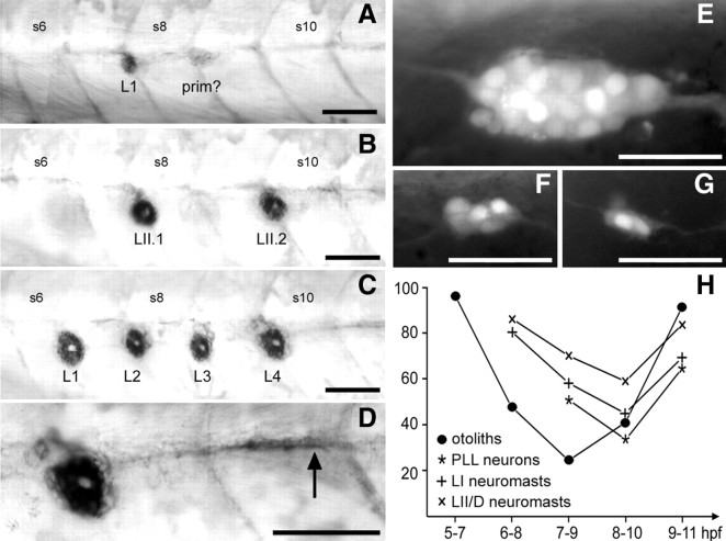
Effect of DEAB treatment on the PLL. A, An extreme case in which only one neuromast, L1, has formed at 4 dpf. Posterior to L1 is an immobilized primordium (prim?); it is not possible to decide whether this corresponds to primI or primII. B, A case in which LI neuromasts, but not LII, are completely absent. LI and LII neuromasts can be distinguished based on the central, unlabeled region that is oriented along the anteroposterior axis in LI neuromasts and along the dorsoventral axis in LII (Nuñez et al., 2009). C, A case in which LII neuromasts, but not LI, are absent. D, Higher magnification of the array shown in C, to illustrate the arrest of interneuromast cells shortly after the last deposited neuromast (arrow). E, Normal pattern of neurons in a 28–30 hpf nbt:dsred embryo, with 21 ± 2 neurons decorated by the fluorescent marker DsRed. F, G, Reduced ganglia after DEAB treatment. H, Time course of the DEAB effect. For each period of treatment (abscissa), the experimental data of Table 1 are expressed as percentage of the control. +, LI neuromasts; ×, LII/D neuromasts; *, neurons; •, otoliths. Scale bars, 50 μm. S6, S8, and S10, Somite numbers.

