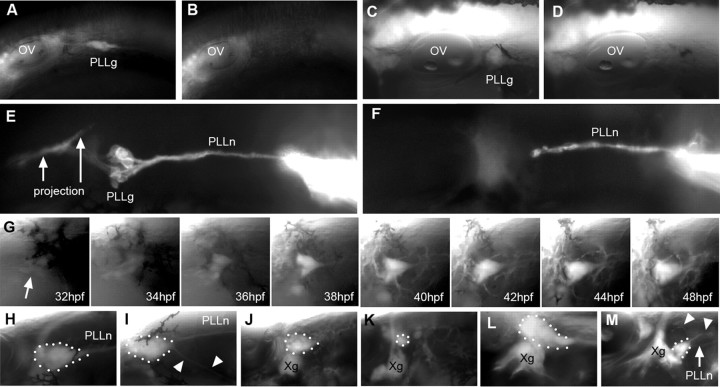
Figure 6.
Regeneration of the PLL ganglion. A, B, PLL ganglion at 24 hpf just before ablation (A) and 2 h later (B). C, D, PLL ganglion at 48 hpf just before (C) and 2 h after ablation (D). E, I, DiI labeling of the PLL nerve in a control embryo (E) and 2 h after ablation of the ganglion at 36 hpf. The PLL projection in the hindbrain is arrowed. G, After ablation at 24 hpf, a successive view of the regenerating ganglion at different times. Panels from 34 to 44 hpf are frames from a time-lapse movie taken at one z-stack every 15 min. The arrow points to the first sign of fluorescence in the region of the ablated ganglion. H–M, Regenerated ganglia 1 d after ablation at 24 hpf (H, I), at 36 hpf (J), at 38 hpf (K), and at 40 hpf (M; L is the control side of the same embryo). Arrowheads in I and M indicate ectopic nerves arising from the regenerated ganglion. Anterior is to the left in all panels except L, which has been inverted along the horizontal axis to facilitate comparison. OV, Otic vesicle; PLLg, PLL ganglion; PLLn, pLL nerve; Xg, vagal ganglion.

