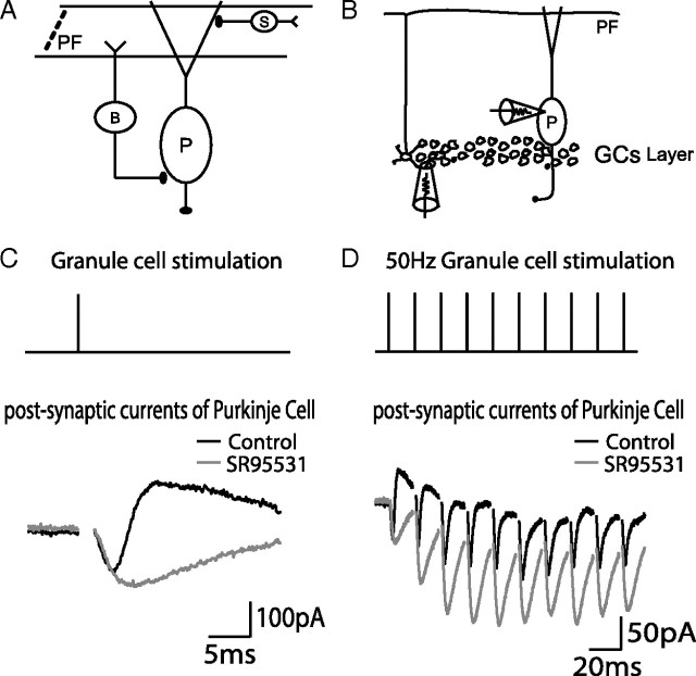
Figure 1.
FFI circuits activated by granule cell stimulation. A, An illustration of somatic and dendritic FFI by PF → BC → PC pathway and PF → SC → PC pathway. B, A diagram showing granule cell extracellular stimulation and somatic patch-clamp recording. C, A single extracellular stimulation applied to the granule cell layer elicited a PSC recorded from the Purkinje cell soma. An excitatory current (negative) is followed by an inhibitory current (positive) in the control condition (black). The GABAA receptor antagonist SR 95531 blocks the inhibitory current (gray). Recorded cells were voltage clamped at −55 mV. Each trace is an average of 10 sweeps, and stimulus artifacts are removed from PSC traces. D, Trains of stimulation pulses were applied to the granule cell layer. An example of a 50 Hz train and its responses are shown in a similar way to C. B, Basket cell; P, Purkinje cell; S, Stellate cell.

