Figure 3.

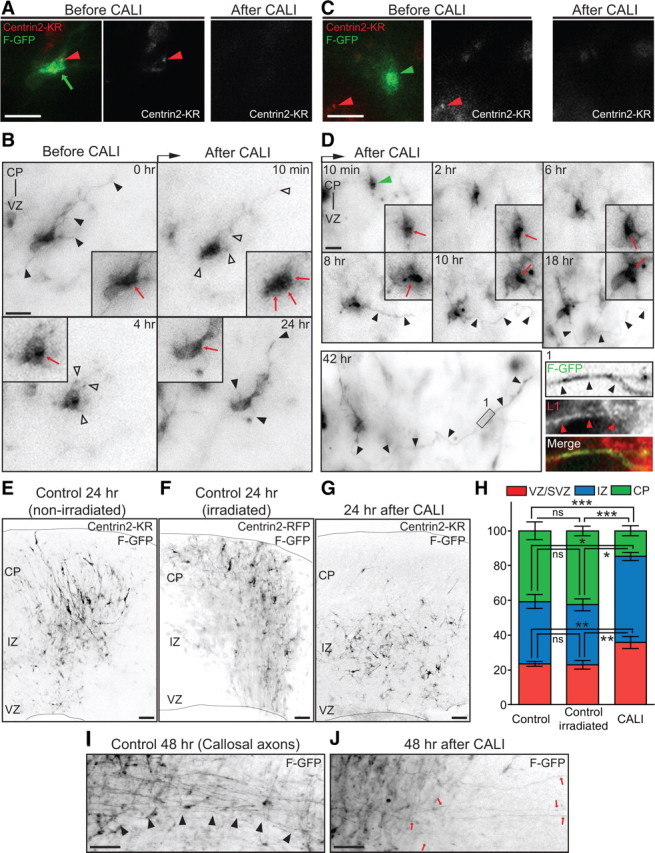
Light-induced inactivation of Centrin2 disrupts axon formation and neuronal migration in situ. A, Centrin2-KR fusion protein (left panel, red arrowhead) localized with the polarized F-GFP signal (left panel, green arrow). B, Before CALI, the cell displayed several neurites (black arrowheads), and a polarized cytoplasm (inset, red arrow). CALI of Centrin2-KR induces neurite retraction (10 min, 4 h, open arrowheads) and redistribution of the polarized F-GFP signal (10 min, inset, red arrows). Eventually, neurites regrew (24 h, black arrowheads) and the cytoplasm repolarized (4 h, 24 h, inset, red arrow). C, Control cells (left panel, green arrowhead) did not express Centrin2-KR (left panel, red arrowhead) but were irradiated with green light. D, The same cell as C (green arrowhead) developed an axon (8 h-42 h, black arrowheads) and markedly polarized cytoplasm (8–18 h, inset, red arrow). The neurite is L1 positive (left bottom, Inset 1 in 42 h, black and red arrowheads). E, F, 24 h after CALI, control slices show labeled cells migrating toward the CP. G, 24 h after CALI, irradiated slices have the majority of the labeled cells in the IZ. H, Quantification of cell distribution in control and irradiated slices 24 h after CALI (mean ± SEM; CP: ***p < 0.0001; IZ: *p = 0.0109; VZ: **p = 0.0043 by one-way ANOVA). I, 48 h after CALI, control slices have a robust callosal axonal tract (black arrowheads). J, 48 h after CALI, treated slices have few F-GFP-positive callosal axons (red arrows). Scale bars: A–D, 10 μm; E–G, I, J, 200 μm.
