
Figure 4.
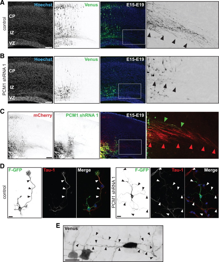
PCM-1 downregulation disrupts axon formation and neuronal migration. A, In utero electroporation of control shRNA at E15 labels transfected cells that form a callosal axonal tract (inset, black arrowheads) at E19. B, PCM-1 shRNA expression resulted in migration defect (middle panel) and failure to form a callosal axonal tract (inset, black arrowheads). C, The cell-autonomous phenotype of PCM-1 shRNA was demonstrated by sequential electroporation of mCherry and PCM-1 shRNA/Venus. Cells with only mCherry expression migrate toward the CP and project callosal axons. PCM-1 shRNA-transfected cells failed to migrate (middle panel) and have limited callosal axonal projections (right inset, green arrowheads). D, PCM-1 downregulation inhibits axon formation in vitro. Polarized control neuron extending a long neurite with typical Tau-1 gradient (black and white arrowheads). PCM-1 downregulation disrupts axon formation but cells have several long (> 40 mm) and thin Tau-1 negative neurites (black and white arrowheads). E, Multipolar cells in the IZ displayed several long and thin neurites after PCM-1 shRNA. Scale bars: A–C, 200 μm; D, E, 10 μm.