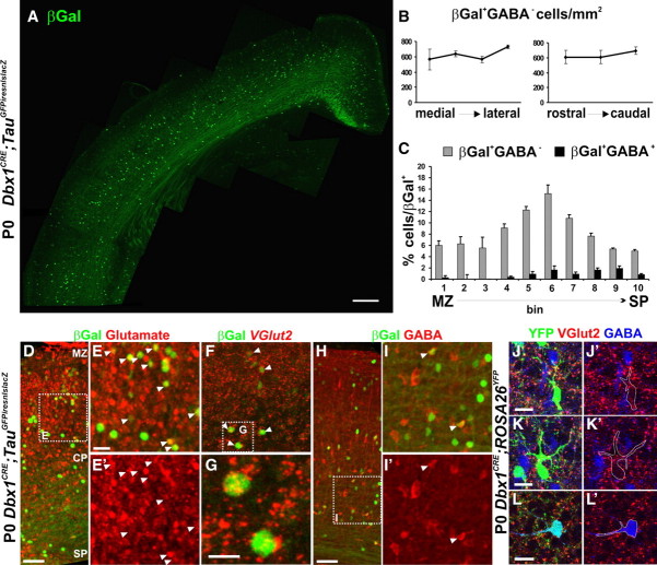
Figure 1.
Distribution and neurochemical properties of Dbx1-derived neurons in the cortical plate. A, Immunohistochemistry for βGal on coronal sections of P0 Dbx1CRE;TauGFPiresnlslacZ animals shows the distribution of Dbx1-derived neurons in the neocortex. B, Graphs show the homogeneous distribution of βGal+GABA− neurons per square millimeter along the mediolateral and rostrocaudal axis (see Materials and Methods). C, Histogram shows the distribution of βGal+GABA− and βGal+GABA+ neurons relative to total number of βGal+ neurons (in percentage) in the dorsolateral neocortex divided in 10 bins in its radial dimension. Results are expressed as mean ± SEM. D–I′, Immunohistochemistry for βGal and glutamate (D–E′) or GABA (H–I′) on coronal sections of P0 Dbx1CRE;TauGFPiresnlslacZ animals. F, G, Fluorescent in situ hybridization for VGlut2 followed by βGal immunostaining on P0 Dbx1CRE;TauGFPiresnlslacZ brains. E, G, I, High magnifications of dashed boxes in D, F, and H, respectively. The vast majority of Dbx1-derived neurons are Glu+ (91.38 ± 3.55%; n = 743 of 813) or VGlut2+ (83.6 ± 1.06%; n = 98 of 116), whereas a small percentage are GABA+ (7.85 ± 0.75%; n = 108 of 1380) or VGlut2− (6.3 ± 0.53%; n = 7 of 116). The white arrowheads show colabeled cells. J–L, Immunohistochemistry for VGlut2, GABA, and YFP on Dbx1CRE;ROSA26YFP animal at P0 shows the neurochemical properties of Dbx1-derived cells in the CP. Many of the YFP+ cells express VGlut2+ but not GABA (J–K′). Conversely, the YFP+GABA+ cells are not labeled with VGlut2 (L, L′). Images in J–L′ represent a 1-μm-thick confocal plane. Scale bars: A, 200 μm; D, E, 50 μm; F, J, K, L, 20 μm; G, 10 μm.

