
Figure 2.
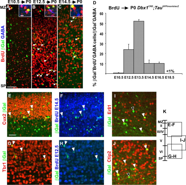
Birthdating of the glutamatergic Dbx1-derived cell population. A–C, BrdU staining at P0 on Dbx1CRE;TauGFPiresnlslacZ animals after a pulse shows that βGal+ neurons born at E10.5 are positioned in the MZ (A); those born at E12.5, in deep layers (B); and those born at E14.5, in more superficial layers (C) (white arrowheads). The dashed boxes in A–C are high magnifications of βGal+BrdU+GABA− neurons used for quantification in D. D, Graph represents the percentage of βGal+BrdU+GABA−/total βGal+GABA− neurons after a pulse of BrdU at different embryonic stages and analyzed at P0. Glutamatergic Dbx1-derived neurons in the CP are mainly born between E11.5 (24.09 ± 5.17%; n = 19 of 78) and E14.5 (10.24 ± 3.07%; n = 54 of 527), with a peak at E12.5 (51.99 ± 1.89%; n = 79 of 152). Results are expressed as mean ± SEM. E–H, βGal+ neurons are predominantly Tbr1+ (G) and Cux2+ (E) when born at E12.5 (G, H) and E14.5 (E, F), respectively (BrdU in blue). I, J, Some of βGal+ neurons positioned in layer V express ER81 (I) or Ctip2 (J). The black and white arrowheads show single- and double-labeled cells, respectively. K, Diagram represents the positions where the respective images have been taken in the dorsolateral cortex of P0 Dbx1CRE;TauGFPiresnlslacZ animals. Scale bars: A, E, 50 μm; A, boxed region, 10 μm.