
Figure 4.
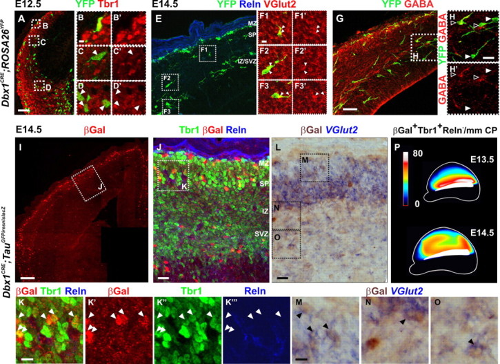
Dbx1-derived glutamatergic neurons present the morphology of tangentially migrating cells and progressively populate the entire pallium by E14.5. A–D′, Immunostaining for YFP and Tbr1 on coronal sections of E12.5 Dbx1CRE;ROSA26YFP animals shows a deep stream of YFP+Tbr1+ cells emerging from the PSB (D, D′) and showing a typical morphology of migrating cells up to the lateral and dorsolateral pallium (C, C′). B and B′ correspond to a Dbx1-derived CR neuron in the MZ (Reln staining not shown). E–H′, Immunostaining for YFP (E–H) and VGlut2 (E–F3′) or GABA (G–H′) on serial coronal sections of E14.5 Dbx1CRE;ROSA26YFP animals labeling either Dbx1-derived glutamatergic neurons (F1–F3′,H,H′, white arrowheads) or inhibitory interneurons (H, H′, black arrowheads). Both populations migrate through the IZ. Some YFP+VGlut2+ neurons are observed invading the CP by radial migration (F1, F1′). Images in F1–F3′ represent a 1-μm-thick confocal plane. I–K‴, Numerous βGal+Tbr1+Reln− cells are detected at rostrodorsal levels in the CP of Dbx1CRE;TauGFPiresnlslacZ animals at E14.5. βGal+Tbr1+Reln+ CR cells position specifically in the MZ. K–K‴, High magnifications of boxed region in J. K′–K‴ show single-channel images. βGal+Tbr1+Reln− cells are distributed in the dorsal CP by E14.5 (white arrowheads). L–O, Immunostaining for βGal after in situ hybridization with a VGlut2 mRNA probe shows double-labeled cells in the IZ, SP, and CP of E14.5 Dbx1CRE;TauGFPiresnlslacZ rostrodorsal cortices. M–O, High magnifications of boxed regions in L. P, Schematic representation of the distribution of βGal+Tbr1+Reln− neurons in the CP of E13.5 and E14.5 Dbx1CRE;TauGFPiresnlslacZ cortices (lateral view) (for values, see supplemental Table 1, available at www.jneurosci.org as supplemental material). Scale bars: I, 100 μm; A, E, G, 50 μm; J, L, 20 μm; F1, K, M, 10 μm.