
Figure 7.
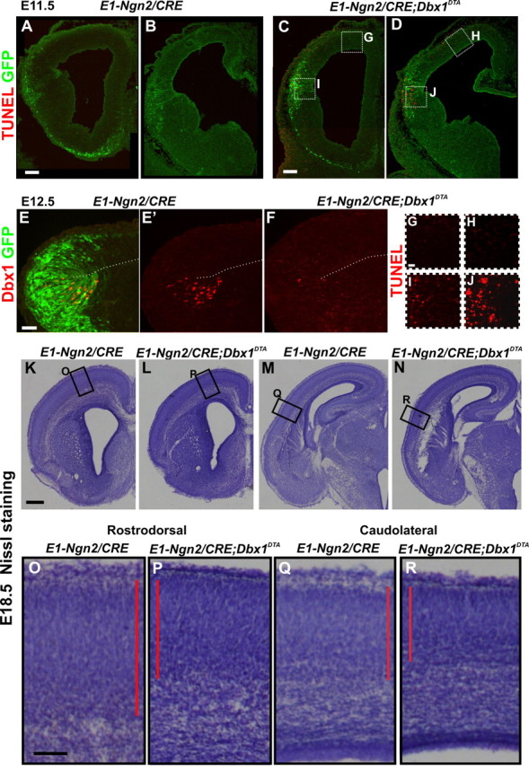
Decrease of cortical plate thickness in E1-Ngn2/CRE;Dbx1DTA animals upon ablation of CP transient cells. A–D, G–J, TUNEL and GFP staining of E11.5 E1-Ngn2/CRE(iresGFP) control (A, B) and E1-Ngn2/CRE;Dbx1DTA (C, D) telencephalons. G–J, High magnifications of boxed regions in C and D. More TUNEL+ cells are detected at the PSB caudally (J) than rostrally (I) in mutants, whereas no cell death is observed in the dorsolateral pallium (G, H) and control telencephalons. E, F, Immunohistochemistry for GFP and Dbx1 on E12.5 embryos showing that the E1-Ngn2/CRE(iresGFP) enhancer is expressed in all Dbx1+ progenitors at the PSB in control animals (E, E′). The number of Dbx1+ progenitors is dramatically reduced in E1-Ngn2/CRE;Dbx1DTA animals at the PSB (compare E′, F). K–R, Nissl staining on coronal sections of E18.5 control (K, M, O, Q) and E1-Ngn2/CRE;Dbx1DTA mutant (L, N, P, R) cortices at rostrodorsal (K, L, O, P) and caudolateral (M, N, Q, R) levels showing a decrease in CP thickness in mutant compared with control animals. Scale bars: K, 250 μm; A, C, 100 μm; E, O, 50 μm; G, 20 μm.