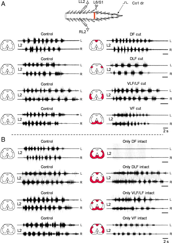
Figure 2.

Effects of funicular lesions on the lumbar rhythm produced by SCA stimulation. A, Recordings from the left and right L2 ventral root in 4 different experiments during stimulation of the Co1 dorsal root before (Control), and after bilateral interruption of the DF (DF cut), DLF (DLF cut), VLF/LF (VLF/LF cut) and VF (VF cut). The lesioned funiculi are shown in red in the schematic cross sections (left to each set of traces). Stimulus trains (control and the respective postlesion recordings), DF cut: 60 pulse, 4 Hz, 1.7T; DLF cut: 50 pulse, 3.3 Hz, 1.2T; VLF/LF cut: 50 pulse, 4 Hz, 1.25T; VF cut: 20 pulse, 1.3 Hz at 1.1T Note that the VLF lesions included the LF. Therefore these lesions are referred to as VLF/LF lesions throughout the present study. B, Recordings from the left and right L2 ventral root in 4 different experiments during stimulation of the Co1 dorsal root before (Control), and after bilateral interruption of the VF, VLF/LF and DLF (only DF intact), the VF, VLF/LF and DF (only DLF intact), the VF, DLF and DF (only VLF/LF intact) and the VLF/LF, DLF and DF (only VF intact). The lesioned funiculi are shown in red in the schematic cross sections (left to each set of traces). Stimulus trains: only DF intact: 50 pulse, 4 Hz, 1.7T (control) and 8.3T (postlesion); only DLF intact 40 and 50 pulse (control and postlesions), 3.3 Hz, 1.9T; only VLF/LF intact: 20 pulse, 1.3 Hz, 1.1T (control), 30 pulse 2 Hz, 1.9T (postlesions); only VF intact: 30 pulse, 2 Hz, 1.2T. Calibration bars (A, B), 2 s.
