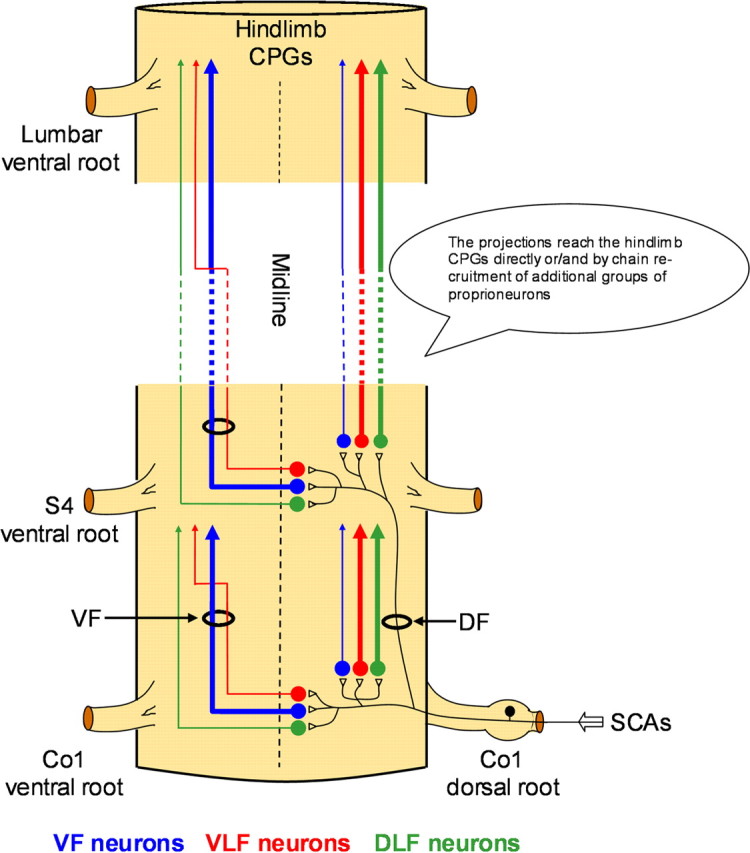
Figure 9.

The pathways involved in activation of the hindlimb CPGs by SCA stimulation. Schematic ventral view of the spinal cord illustrates the suggested functional organization of the pathways activated by SCA stimulation to produce rhythmic activity in the limb innervating segments of the spinal cord. Sacrocaudal afferents (SCAs) entering the spinal cord through the sacral dorsal roots (Co1 in this example) activate segmental sacral neurons that project rostrally via the VF (blue), VLF (red) and DLF (green) as demonstrated in our lesion experiments (Figs. 2 and 3). Nonsegmental sacral neurons (S4 in this example) with similar projection pattern are activated by the Co1 afferents ascending within the sacral dorsal funiculus (DF), as demonstrated in the experiment shown in Figure 4. The proportions of crossed and uncrossed projections through each funiculus (based on the labeling studies shown in Figs. 6–8) are denoted by the thickness of the illustrated pathways. Thus, the VF projections are mainly crossed and the VLF and DLF are mainly uncrossed. Some of the crossed VLF projections join the VLF after traveling through part of the sacral VF as demonstrated in Figure 7. The axons of the activated sacral neurons reach the hindlimb pattern generators either directly (see Fig. 5A) or/and by chain recruitment of groups of proprioneurons (see Fig. 5B). Projections of VF neurons can activate other groups of VF, as well as VLF and DLF proprioneurons (see Fig. 5B and text).
