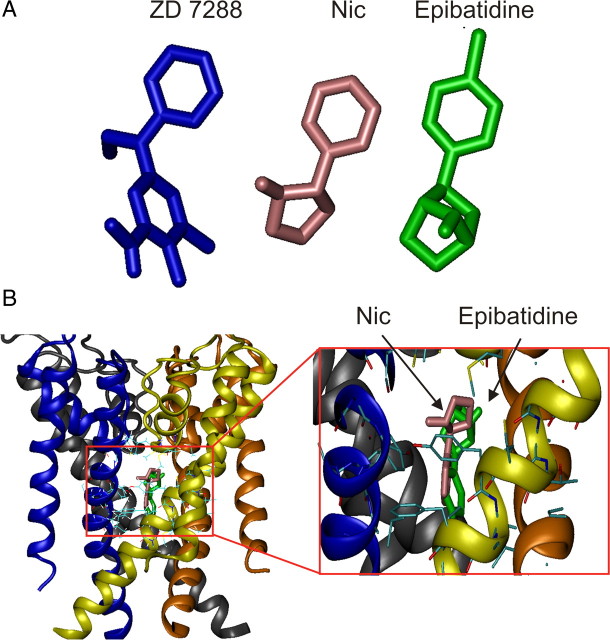
Figure 11.
Putative binding site of nicotine and epibatidine into the inner pore of HCN channels. A, Three-dimensional structures of ZD 7288, nicotine (Nic), and epibatidine, respectively. B, Model of the tetrameric HCN channel pore structure with nicotine (pink arrow) and epibatidine (green arrow), docked in the internal cavity of the channel. Surrounding amino acids are shown in stick representation. For clarity, the four monomers are shown in different colors.

