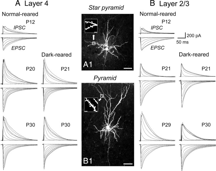
Figure 1.
Representative traces of eIPSC and eEPSC at different postnatal days and in the different rearing conditions. A, B, Examples of responses evoked by stimulation at a series of increasing intensity in a star pyramid cell in layer 4 (A) and a pyramidal cell in layer 2/3 (B) of the mouse visual cortex at the indicated postnatal days. Traces in the left and right columns in A and B show those obtained from normally reared and dark-reared mice, respectively. A1, B1, Images of a star pyramidal cell in layer 4 and a pyramidal cell in layer 2/3, stained with biocytin. Insets in A1 and B1 show magnified images of the boxed area in which dendritic spines are visible. Scale bars: A1, B1, 50 μm; insets, 5 μm..

