
Figure 1.
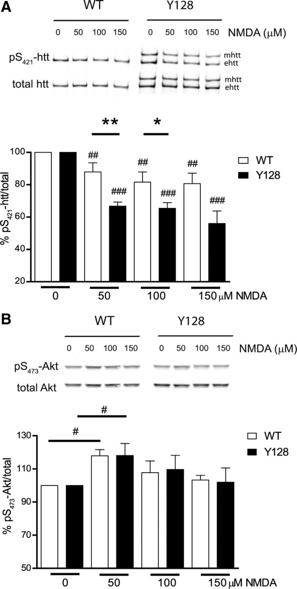
YAC128 primary neurons show loss of phosphorylation of pS421-htt after NMDA-induced excitotoxicity. A, Cortical neurons were treated with varying doses of NMDA, 30 μm glycine for 10 min at 12 DIV. After drug removal, neurons were cultured for 15 min, and protein lysates were either processed for immunoprecipitation of htt or analyzed as described in B. Immunoprecipitated htt was separated by Western blot and membranes were probed with Abs against pS421-htt and total htt (mAb 2166) followed by probing with red- (goat anti-rabbit Alexa Fluor 680) and green-labeled (goat anti-mouse IRDye-800-CW) secondary Abs. Quantification of immunoreactivity was performed with a LI-COR system using Odyssey software. The ratio between pS421-htt and total htt was determined and expressed relative to controls for each condition and genotype in each of eight independent experiments. Relative levels of phosphorylation for mutant htt (mhtt) and endogenous htt (ehtt) were pooled. *p < 0.05, **p < 0.005 compared with WT between genotypes, by t test; ##p < 0.005, ###p < 0.0005 compared with control within genotype, by paired t test. Error bars denote ±SEM. B, Protein lysates, prepared as described in A, were separated by Western blot and probed with Abs against pS473-Akt and total Akt. The ratio between pS473-Akt and total Akt was determined and expressed relative to controls. Ratios for each condition and genotype in each of five independent experiments were determined. #p < 0.05 compared with controls within genotypes, by paired t test. Error bars denote ±SEM.