Figure 1.

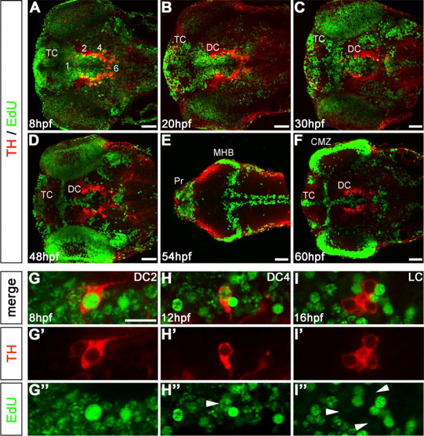
Birth dating of catecholaminergic neurons by pulsed EdU incorporation. A–F, Embryos were labeled with EdU at defined developmental stages, as indicated in panels, and analyzed at 75 hpf by TH immunohistochemistry (red) and EdU Click-iT Alexa 488 label (green). A, Pulse at 8 hpf (75% epiboly) shows most of the neural precursors were still proliferating. B, C, Pulse label at 20 and 30 hpf, Proliferative areas became more restricted. D, Pulse at 48 hpf, EdU labeling retracted to the ventricular proliferative zones (VZ). E, Pulse label at 54 hpf, Dorsal focal planes only: proliferative zones of the midbrain hindbrain boundary (MHB), rhombencephalic VZ, and many cells in the pretectum (Pr) were labeled. F, Pulse at 60 hpf, VZ of the diencephalon and telencephalon (DC and TC, respectively), and retinal ciliary marginal zone (CMZ) were labeled. G–I, EdU label evaluation. G–G″, DC2 THir cell with a bright, homogenous EdU nuclear label considered as a cell that became postmitotic soon after the incorporation of EdU. H–H″, DC4 THir cell with a spotted green nuclear label, considered as a cell that passed through several more cell cycles after incorporating EdU (white arrowhead). I–I″, LC THir cells without green-labeled nuclei (white arrowheads), considered either as cells already postmitotic at the time point of EdU incubation or as cells that passed through so many cell divisions until 75 hpf, that the EdU was not detectable anymore. Given that time windows of CA differentiation are known based on the onset of TH immunoreactivity, both options could be distinguished. A–F, Z-projections of every fifth image plane to show all relevant THir clusters in one image; dorsal views, anterior left. Scale bars: A–F, 50 μm; (in G) G–I″, 20 μm.
