Figure 1.
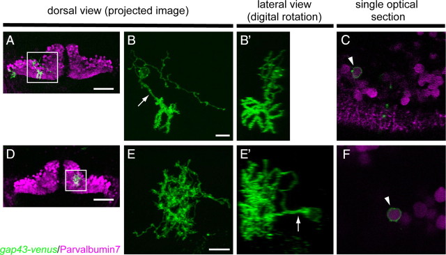
Dendritic morphology of zebrafish Purkinje cells. A, D, Dorsal view images of the cerebellum of 8 dpf larvae that were injected with aldoca:gap43-Venus plasmid and mosaically expressed gap43-Venus in the Purkinje cells. Two Purkinje cells in A and one in D expressed gap43-Venus (green). All the Purkinje cells were labeled with an anti-parvalbumin7 antibody (magenta). Purkinje cells surrounded by boxes were subjected to observation at a higher resolution (B, B′, C, E, E′, F). Scale bars, 20 μm. B, B′, Dendritic morphology of a relatively posterolateral Purkinje cell labeled with gap43-Venus, boxed in A. B, Projected image of serial confocal optical sections taken from the dorsal side. The Purkinje cell extended a single primary dendrite (arrow) posteriorly. Dense dendritic spines were observed at the dendritic terminals. B′, Lateral view constructed digitally. Dendritic arbors expanded along the dorsoventral axis. Scale bar, 10 μm. E, E′, Dendritic morphology of an anteromedial Purkinje cell, boxed in D. E, Dorsal view with a projected image. E′, Lateral view. The Purkinje cell extended a single primary dendrite dorsally, and the dendritic arbors were randomly distributed. C, F, Single optical sections at the level of the soma of the posterolateral (C) and anteromedial (F) Purkinje cells shown in B and E, respectively. The gap43-Venus-expressing Purkinje cells were positive for parvalbumin7 (magenta). Scale bar, 10 μm.

