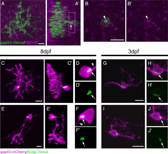
Figure 3.
Golgi apparatus is exclusively localized to the root of the primary dendrite, and this localization precedes the primary dendrite specification. A–B′, A Purkinje cell expressing gap43-Venus (green) in an 8 dpf larva that was immunostained with the GM130 antibody (magenta). A, A′, A dorsal view projected image (A) and a lateral view constructed digitally (A′). The GM130-positive Golgi was localized to the root of the primary dendrite (small arrow). B, B′, Higher-magnification views of the projected image of optical sections corresponding to the boxed area in A′. The GM130 signal was only observed at the root of the primary dendrite (arrowheads). C–J′, Purkinje cells in larvae expressing the aldoca:gap43-mCherry-PTV1–2A-Golgi-Venus plasmid. Using the 2A peptide system, the cellular morphology was visualized with gap43-mCherry (magenta), and the Golgi apparatus was visualized with Golgi-Venus (green). C–E′, Purkinje cells in 8 dpf larvae. Dorsal (C, E, projected images) and lateral (C′, E′, constructed digitally) views of anteromedial (C, C′) and posterolateral (E, E′) Purkinje cells. D, D′, and F, F′ are high-magnification views of a single optical section of the Purkinje cells in C and E, respectively. The Golgi apparatuses that were visualized with Golgi-Venus (arrows) were exclusively localized to the root of the primary dendrites (arrowheads). G, I, Dorsal view projected images of Purkinje cells in 3 dpf larvae. H, H′ and J, J′ are high-magnification views of a single optical section in the Purkinje cells in G and I, respectively. At this stage, Purkinje cells had multiple primary dendrites and did not exhibit morphological polarity, but the Golgi apparatus was already localized to a restricted area (arrows). Scale bars, 10 μm.

