. 2019 Jun 26;8:e45282. doi: 10.7554/eLife.45282

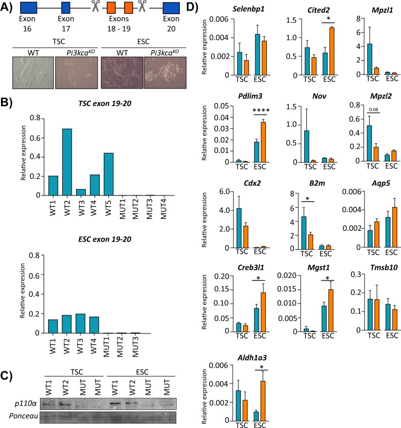
Figure 7. PI3K p110α differentially affects the expression of genes in the trophoblast and fetal cell lineages.

(A) Details of CRISPR/Cas9 design to delete exons 18–19 of Pik3ca in trophoblast or embryonic stem cells (TSC and ESC, respectively). The deletion in TSC (trophoblast stem cells) and ESC (embryonic stem cells) was confirmed by (B) qRT-PCR analysis of exons 18–19 of Pik3ca and (C) by Western blotting for p110α. (D) The expression of candidate genes in Pi3kca wildtype (WT) and mutant (Mut) TSC (n = 5 and n = 4, respectively) and ESC (n = 4 and n = 3, respectively). Data in (D) are normalized against housekeeping Sdha. *p<0.05, ****p<0.0001, two-ways ANOVA analysis followed by Fisher post hoc test. Data presented as means ± SEM.

Figure 7—source data 1. Functional predictions for the candidate genes enriched in trophoblast and embryonic stem cells cells (TSC and ESC, respectively).
DOI: 10.7554/eLife.45282.025
Figure 7—source data 2. Primer sequence for RT-qPCR, PCR screening and CRISPR gRNAs target.
DOI: 10.7554/eLife.45282.026

Figure 7.

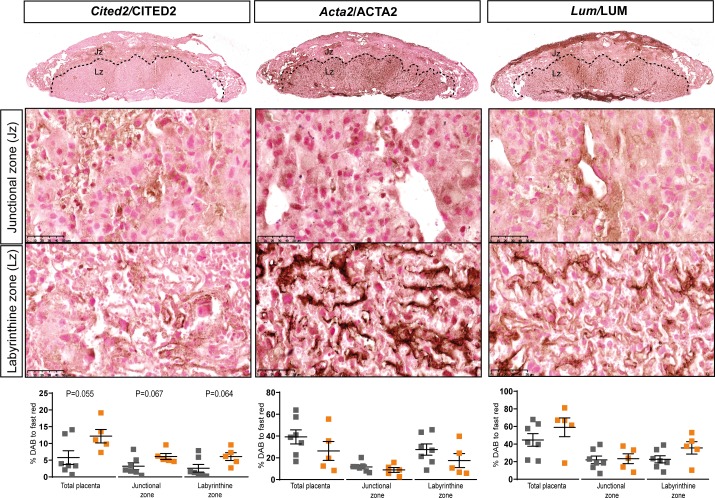
Figure 7—figure supplement 1. Localisation and abundance of CITED2, ACTA2 and LUM identified to be dysregulated at the gene level, in Hom-P versus Het-U placentas on day 19 of pregnancy.

Figure 7—figure supplement 1.

Data presented as individual values with means ± SEM shown. Data are from n = 7 for Het-U and n = 5 for Hom-P and were analysed by unpaired t test.
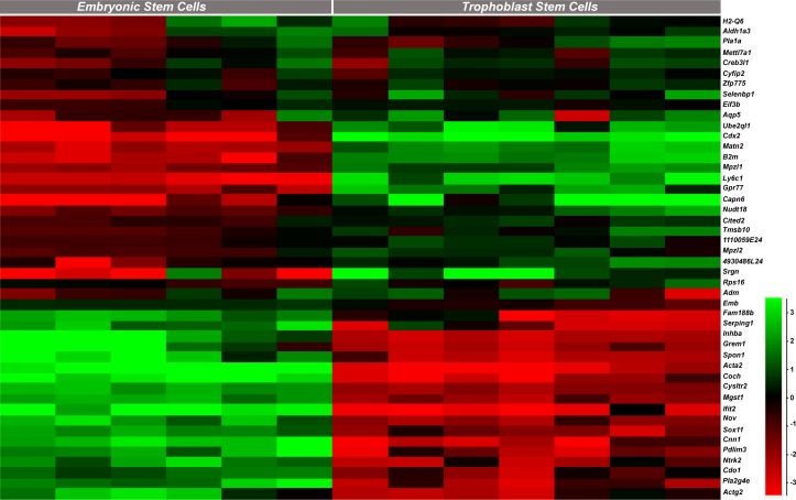
Figure 7—figure supplement 2. Expression profiles of the genes identified as dysregulated in Hom-P versus Het-U placentas in trophoblast stem cells (TS cells) and embryonic stem cells (ES cells), as determined by analysis of existing RNA-seq datasets (Chrysanthou et al., 2018; Latos et al., 2015).

Figure 7—figure supplement 2.