. 2019 Jul 16;8:e47815. doi: 10.7554/eLife.47815

Figure 2. Carbon exchange between N.oceanica and M. elongata AG77.

(A) Carbon (C) transfer from [14C]-sodium bicarbonate (NaHCO3)-labeled N. oceanica (Noc) cells to M. elongata AG77 (Mel AG77, left panel) or from [14C]-glucose-labeled AG77 to Noc cells (right panel) after 7-d co-culture in flasks (with physical contact). Radioactivity of 14C-carbon was determined with a scintillation counter (dpm, radioactive disintegrations per minute) and then normalized to the dry weight of samples (dpm/mg biomass). Free Noc, unbound Noc cells in the supernatant; attached, Noc cells separated from AG77-Noc aggregates by algal cell wall digestion and mesh filtration; FAAs, free amino acids; soluble compounds, supernatant after acetone precipitation of proteins extracted by SDS buffer. Data are presented as the average of three biological replicates with standard deviation (Means ± SD, n = 3). (B) 14C-carbon transfer between Noc and AG77 without physical contact. Algae and fungi were incubated in cell-culture plates with filter-bottom inserts (pore size of 0.4 μm) which separate Noc cells and AG77 mycelium from each other but allow metabolite exchange during co-culture. Error bars indicate SD of three biological replicates (n = 3). (C and D) Relative abundance of 14C-carbon radioactivity in recipient cells compared to 14C-labeled donor cells after 7-d co-culture. (C) AG77 relative to [14C]-NaHCO3-Noc (100%). (D) Noc relative to [14C]-glucose-labeled AG77 (100%). Physical contact, living 14C-labeled cells added to unlabeled cells for co-cultivation in flasks; no contact, samples grown separately in plates with inserts; heat-killed 14C-cells, 14C-labeled Noc or AG77 killed by heat treatment at 65°C for 15 min before the addition to unlabeled cells in flasks. Free, unbound Noc cells in the supernatant; Att, Noc cells attached to AG77 (isolated by algal cell wall digestion and mesh filtration); Total, Noc cells grown separately from AG77 in plates and inserts. Error bars indicate SD of three biological replicates (n = 3). (E) Nitrogen (N) exchange between N. oceanica (Noc) and M. elongata AG77 examined by 15N-labeling experiments. [15N]-potassium nitrate-labeled Noc cells or [15N]-ammonium chloride-labeled AG77 were added to unlabeled AG77 or Noc cells, respectively, for 7-day co-culture in flasks (physical contact) or cell-culture plates with inserts (no physical contact). Algae and fungi were separated and weighed (dry biomass) after the co-culture, and their isotopic composition in Atom% 15N [15N/(15N+14N)100%] and N content (%N) were determined using an elemental analyzer interfaced to an Elementar Isoprime mass spectrometer following standard protocols. The N uptake rate of 15N-Noc-derived N (15N) by AG77 from and that of 15N-AG77-derived N by Noc cells (15N) were calculated based on the Atom% 15N, %N and biomass. C, chloroplast; N, nucleus; Nu, nucleolus; M, mitochondrion; V, vacuole; L, lipid droplet. Values are the average of three biological repeats.
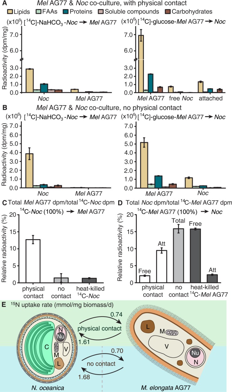
Figure 2.

Figure 2—figure supplement 1. Carbon transfer between N.oceanica and M. elongata AG77 with physical contact.

Figure 2—figure supplement 1.

Carbon transfer between N. oceanica and M. elongata AG77 with physical contact. Separation of N. oceanica and M. elongata AG77 after co-culture and exchange of radioactive carbon ([14C]) between N. oceanica and M. elongata during co-culture. (A) Aggregates of N. oceanica (Noc) cells and AG77 mycelium after 7-day co-culture. (B) Isolated AG77 mycelium from AG77-Noc aggregates after the digestion of hemicellulase and driselase and vortex washes. (C) Isolated Noc cells by mesh filtrations using Accu-Mesh PW200 (yellow mesh, lower right) and then SEFAR NITEX 03-25/14 (white mesh, lower right, mesh opening 25 μm). (D) PCR amplification to examine Noc contamination in isolated AG77 mycelium using primers specific for the gene encoding Aureochrome 4 (AUREO4) in Noc. Primers specific for the fungal gene encoding translation elongation factor EF1α (EF1α) were used as positive controls for fungal tissue. Samples #1 to #3 are biological replicates of isolated fungal mycelium. (E) PCR test for fungal contamination in isolated Noc cells with primers specific for fungal genes encoding EF1α and RNA polymerase RPB1 (RPB1). AUREO4 primers were used as positive controls for the algal cells. Samples #1 and #2 are collected after filtration with PW200 and PW200/NITEX meshes, respectively. (F) Co-culture of N. oceanica (Noc) and M. elongata AG77 in flasks. [14C]-labeled cells were added to unlabeled cells and co-cultured for 7 days; left flask, [14C]-sodium bicarbonate (NaHCO3)-labeled Noc; middle flask, [14C]-glucose-labeled AG77; right flask, [14C]-sodium acetate-labeled AG77. (G) Normalized radioactivity of 14C with respect to the dry weight (dpm/mg). Dpm, radioactive disintegrations per minute; free Noc, unbound cells harvested from the supernatant; attached, Noc cells separated from Noc-AG77 aggregates; FAAs, free amino acids; soluble compounds, supernatant after acetone precipitation of SDS-protein extraction. Values are shown as the average of three biological replicates with standard deviation (Means ± SD, n = 3).
Figure 2—figure supplement 2. Carbon and nitrogen exchange between N.oceanica and M. elongata AG77 without physical contact.

Figure 2—figure supplement 2.

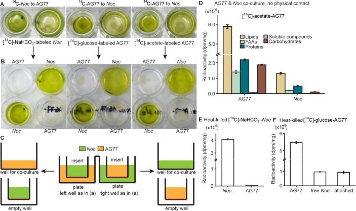
Carbon and nitrogen exchange between N. oceanica and M. elongata AG77 without physical contact. 14C exchange between N. oceanica and M. elongata AG77 without physical contact using co-culture of N. oceanica (Noc) and M. elongata AG77 in 6-well plates with filter-bottom inserts. (A) Co-cultivation without physical contact. The hydrophilic polytetrafluoroethylene filter (pore size of 0.4 μm) at the bottom of the inserts separates Noc and AG77 during co-culture but allows metabolic exchange between the plate well and insert. [14C]-sodium bicarbonate (NaHCO3)-labeled Noc cells were grown in the plate well or insert while recipient AG77 was grown in the insert or plate well, respectively. Similar incubation conditions were used for [14C]-glucose- or [14C]-sodium acetate-labeled AG77 and recipient Noc. (B) After 7-day co-culture, the inserts were moved to the adjacent empty wells (bottom) for harvesting samples. There was no cross contamination observed between Noc and AG77 samples. (C) Side-view diagram of alga-fungus co-culture (A) and sample harvesting (B) with insert and plate. (D) Detection of 14C transfer from [14C]-sodium acetate-labeled AG77 to recipient Noc. 14C radioactivity (dpm, radioactive disintegrations per minute) was normalized to the dry weight (dpm/mg). FAAs, free amino acids; soluble compounds, supernatant after acetone precipitation of SDS-protein extraction. Values are shown as the average of three biological replicates with standard deviation. (E) 14C transfer between N. oceanica and M. elongata AG77 co-cultured in flasks with physical contact using heat-killed 14C-cells. [14C]-sodium bicarbonate (NaHCO3)-labeled N. oceanica (Noc) cells were killed by incubating them in a water bath at 65°C for 15 min before co-culture with recipient M. elongata AG77 in flasks. 14C radioactivity (dpm, radioactive disintegrations per minute) was measured after separation of Noc and AG77 and then normalized to the dry weight (dpm/mg). Error bars indicate SD, n = 3. (F) Heat-killed [14C]-glucose-labeled AG77 (65°C, 15 min) was added into unlabeled Noc culture and co-cultivated for 7 days in flasks. Normalized radioactivity of 14C is shown as the average of three biological repeats. Free Noc, unbound cells harvested from the supernatant; attached, Noc cells separated from Noc-AG77 aggregates. Nitrogen (N) exchange between N. oceanica (Noc) and M. elongata AG77 examined by 15N-labeling experiments.
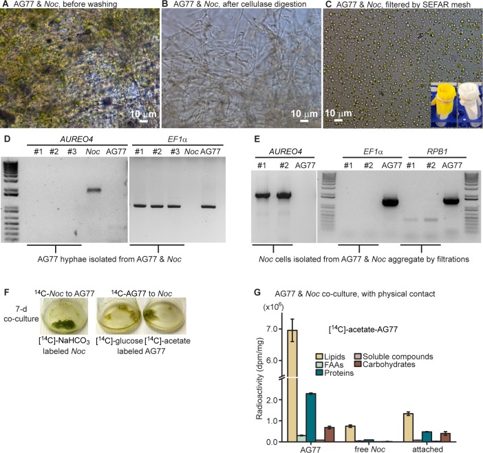
Figure 2—figure supplement 3. Viability of N.oceanica and M. elongata AG77 during 7-d co-culture.

Figure 2—figure supplement 3.

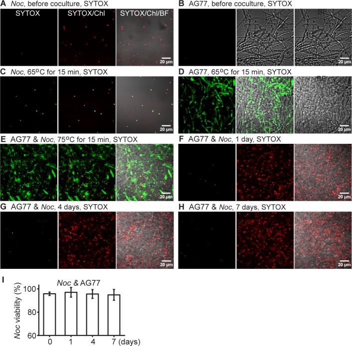
Viability of N. oceanica and M. elongata AG77 during 7-d co-culture. (A–E) Detection of dead cells in N. oceanica (Noc) mid-log phase culture (A) and M. elongata AG77 grown in PDB medium (B) using SYTOX Green staining. In terms of SYTOX Green-positive controls, Noc (C), AG77 (D) and AG77-Noc aggregates (E) were killed by high temperature in a water bath (65 or 75°C for 15 min) and stained with SYTOX Green. Green fluorescence, SYTOX Green (SYTOX) indicating dead cells; red, Noc chlorophyll fluorescence (Chl); BF, bright field. (F–H) SYTOX Green staining of Noc cells when co-cultured with AG77 for 1 d (F), 4 d (G) and 7 d (H). (I) Viability of Noc cells shown in (F–H) with the 0-d control. Results are calculated from ~2000 cells of six biological repeats with ImageJ. Values are shown as the average of six biological replicates with standard deviation.
Figure 2—figure supplement 4. Nitrogen exchange between N.oceanica and M. elongata AG77.

Figure 2—figure supplement 4.

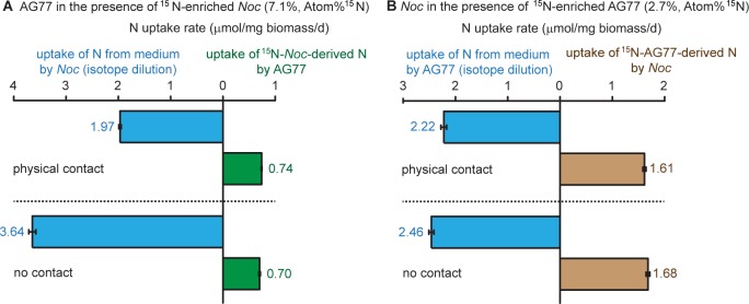
Nitrogen exchange between N. oceanica (Noc) and M. elongata. (A) 15N-potassium nitrate-labeled Noc cells 7.1%, Atom% 15N, 15N/(15N+14N)100% were added to unlabeled AG77 for 7-day co-culture in flasks (physical contact, top) or cell-culture plates with inserts (no physical contact, bottom). Algal and fungal cells were separated and weighed (dry biomass) after the co-culture, and their isotopic composition Atom% 15N [15N/(15N+14N)100%] and N content (%N) were determined using an elemental analyzer interfaced to an Elementar Isoprime mass spectrometer following a standard protocol. The N uptake rates (mmol N/mg biomass/d) of Noc from the medium (medium-N, isotope dilution) and that of AG77 from 15N-Noc-derived N (15N) were calculated based on the Atom% 15N, %N and biomass. Values are shown as the average of three biological replicates with standard deviation, n = 3. (B) Similar analyses were carried out with 15N ammonium chloride-labeled AG77 (2.7%, Atom% 15N) and unlabeled Noc cells to calculate the uptake rate of medium-N by AG77 and that of 15N-AG77-derived N (15N) by Noc cells. Error bars indicate SD, n = 3.