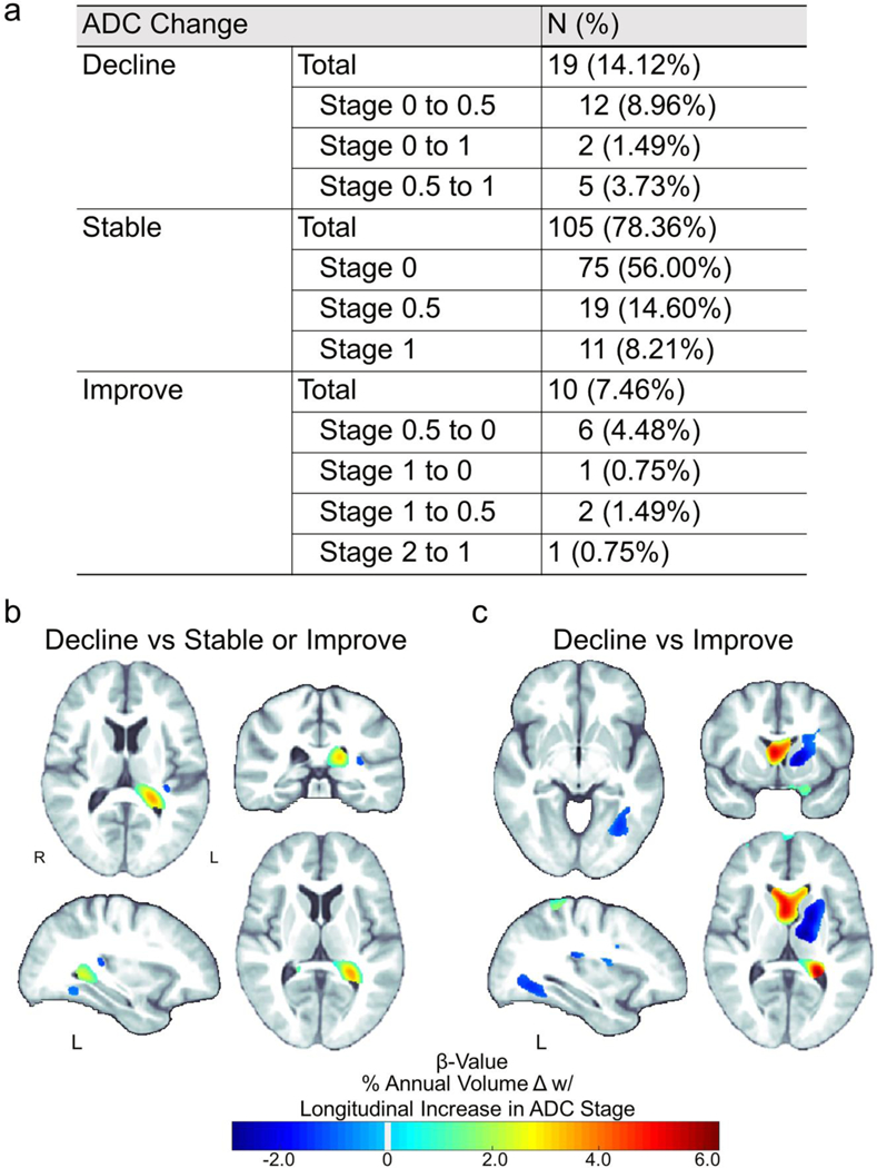
Fig. 3.

Brain tissue loss rates are associated with change in ADC stage. (a) Total number of participants (out of 134) for each type of change in ADC stage between baseline and follow-up assessments. Relative to participants with (b) stable or improved neurocognitive status (no change or decreased ADC stage; N=115) or (c) just improved neurocognitive status (decreased ADC stage; N=10) over time, a decline in neurocognitive status (an increase in ADC stage; N=19) was significantly associated (Pcorrected ≤ 0.05) with greater rates of tissue atrophy (blue) and ventricular expansion (green-red)
