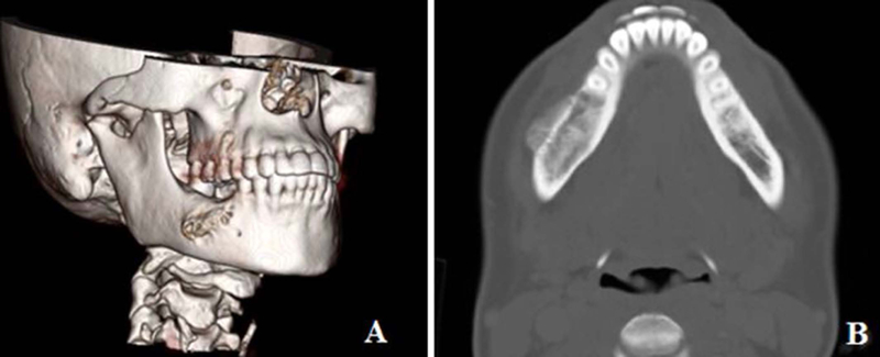
Figure. 4:

3 D reconstruction of the CT scan of the mandible showing only slight expansion of the lateral cortex of the madible (A), and axial view of the CT scan in bone window majority of the tumor being endosteal with slight expansion of the lateral cortex (B).
