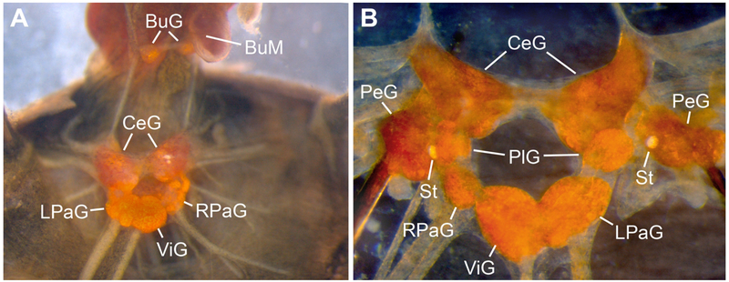
Figure 1.
The central nervous system (CNS) of Biomphalaria alexandrina in various stages of dissection. A) Dorsal perspective of a semi-isolated CNS following removal of the esophagus. The pedal ganglia (PeG) are obscured by the overlying cerebral ganglia (CeG). B) Ventral perspective of an isolated CNS with the pedal commissure cut and pedal ganglia pinned laterally (BuG = buccal ganglia; BuM = buccal mass; LPaG = left parietal ganglion; PlG = pleural ganglia; RPaG = right parietal ganglion; St = statocyst; ViG = visceral ganglion).

