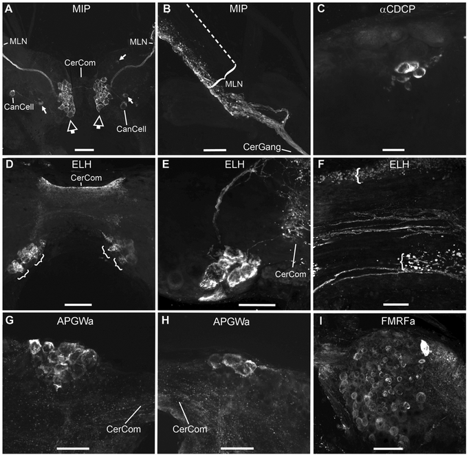
Figure 2.
Neuropeptides in the cerebral ganglia of Biomphalaria alexandrina. A: Molluscan insulin-related peptide (MIP)-like immunoreactivity is located in symmetric clusters of medial neurons (larger, hollow arrows), adjacent to cerebral commissure (CerCom). A bundle of axons projects from each cluster to the median lip nerve (MLN). A single canopy cell (CanCell) was also labelled in each ganglion. On the left, the cell assumed its characteristic lateral position whereas on the right, the cell was located more centrally. A few additional, immunoreactive cells (smaller arrows) are scattered in other regions of the ganglia. B: After the tight bundle of MIP-LIR axons exits the cerebral ganglion (CerGang), it projects to the lateral surface of the MLN where it appears to form a neurohaemal region along its length. The dotted line indicates the location of the faint medial edge of the MLN. C: Antibodies against the α-caudodorsal cell peptide (αCDCP) of Lymnaea label a cluster of small cells near the center of the cerebral ganglion. D: Antibodies against the egg laying hormone (ELH) of Aplysia, reveal two bilateral clusters of large neurons (indicated by brackets) posterior to the cerebral commissure (CerCom). The CerCom itself was also intensely fluorescent. E: A higher magnification image of ELH-positive cells projecting into the adjacent to the CerCom which also contains numerous immunoreactive varicosities. F: Another high magnification image of both intact, ELH-positive axons traversing the CerCom and also numerous varicosities on the anterior and posterior margins of the commissure (indicated by brackets), which appear to constitute another neurohaemal region for peptide release. G: A cluster of large APGWamide-like-immunoreactive cells in the left anterior lobe. H: APGWamide-like-immunoreactive neurons in the right anterior lobe of the same snail as shown in G. I: FMRFamide-like-immunoreactivity in numerous small neurons in the left ventral lobe. All images are oriented to present anterior toward the top of the plate and all images, except for I, show dorsal views. Scale bars equal: 100 μm in A and D; 50 μm in B, E, G and H; 25 μm in C and F; and 20 μm in I.

