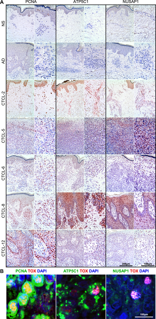
Fig. 4. High numbers of ATP5C1+TOX+, PCNA+TOX+, and NUSAP1+TOX+ T cells accumulate in the skin tumors of patients with advanced-stage CTCL.
(A) Immunohistochemical stain from skin biopsies of normal skin (NS, n=4), atopic dermatitis (AD, n=4), and advanced-stage CTCL (n=5) used in scRNA-seq experiments, each at 200X (left) and 400X (right). (B) Representative examples from 3 patient samples tested of double color immunofluorescence staining for ATP5C1/TOX, PCNA/TOX, and NUSAP1/TOX, as indicated, at 1000X. DAPI stains nuclei.

