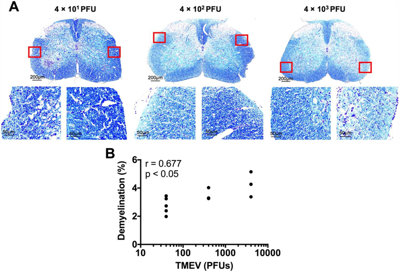
Fig 9.
Spinal cord demyelination increases with the amount of TMEV used to infect the mice. Luxol fast blue was used to stain for myelin. (A) Representative images of spinal cord demyelination with varying TMEV amount, as indicated. (B) Pearson correlation analysis shows significant correlation between quantified demyelination and the amount of TMEV used to infect the mice. (n = at least 3 mice per group and at least two sections per mouse; r = 0.667; p < 0.05).

