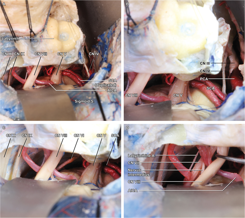
Fig. 4.

Intradural exposure. ( A ) The final exposure is achieved by positioning broad retractors along the interior temporal gyrus and sigmoid sinus/cerebellum. ( B ) Superiorly, the posterior petrosectomy allows visualization to the level of the oculomotor nerve (cranial nerve [CN] III), emerging from the medial midbrain between posterior cerebral artery and superior cerebellar artery. ( C ) Inferiorly, the lower CNs are well demonstrated, as is the abducens nerve (CN VI) deep to CN VII. ( D ) Gentle retraction of the vestibulocochlear nerve (CN VIII) reveals the full course of CN VII, with the nervus intermedius running between them, and the labyrinthine branch of AICA entering the IAC together with the CN VII/VIII complex.
