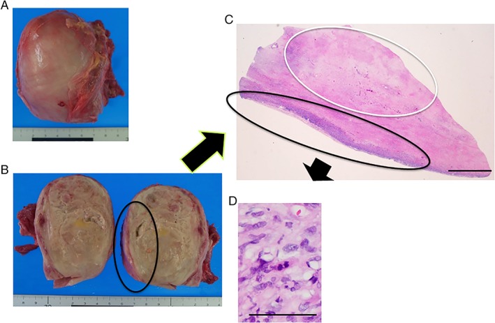
Figure 2.

Pathological findings at autopsy. (A, B) Macroscopic appearance. (C, D) Histological analysis demonstrated glass‐like material inside the neck tumour (necrosis; white circle), which was surrounded by malignant cells (black circle). Haematoxylin and eosin staining, (C) scale bar = 5 mm, (D) scale bar = 100 μm.
