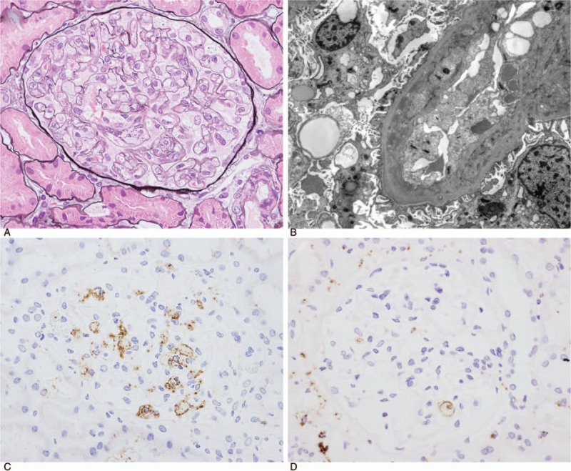
Figure 1.

(A) Light microscopy revealed narrowing of the glomerular capillary lumina owing to diffuse glomerular basement membrane double contours (periodic acid-methenamine-silver [PAM] stain, ×400, high-power field, Case 4). (B) Electron microscopy revealed narrowed glomerular capillary lumina, double contours of the glomerular basement membrane, and partial foot process effacement (Case 5). (C, D) Immunoperoxidase staining for CD68. Glomerular infiltration of CD68-positive cells was observed (C, Case 4; D, Case 5).
