
Figure 1.
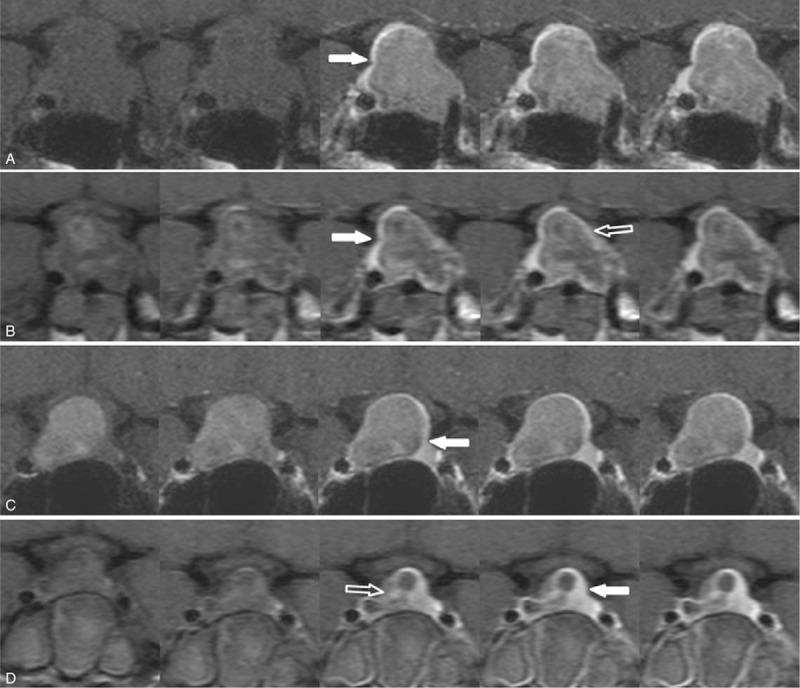
Normal gland on preoperative and postoperative dynamic contrast-enhanced MR images of 2 residual enhancement patterns. Peripheral enhancement pattern (a, b) and nodular enhancement pattern (c, d). On preoperative MRI (a, c), the location of the normal gland (solid arrows) was demonstrated against the pituitary adenoma. Upon immediate postoperative MRI (b, d), the normal gland (solid arrows) and residual enhancing lesion (open arrows) were delineated.MRI = magnetic resonance imaging.