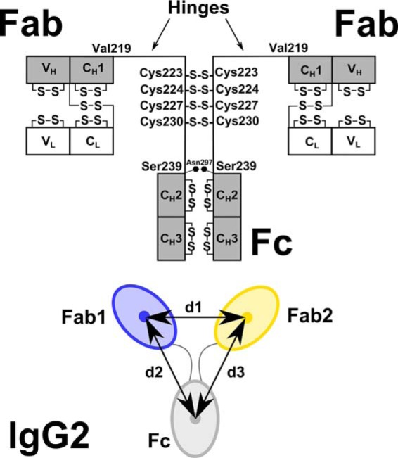
Figure 1.

Human IgG2 domain structure. The two heavy chains each possess VH, CH1, CH2, and CH3 domains, and the two light chains each possess VL and CL domains. The heavy chains are connected by four Cys–Cys disulfide bridges at Cys-223, Cys-224, Cys-227, and Cys-230. There is one N-linked oligosaccharide site at Asn-297 on each of the CH2 domains. The hinge region between the Fab and Fc fragments is composed of 19 residues (ERKCCVECPPCPAPPVAGP) between Val-219 and Ser-239 (EU numbering). Below the black diagram, the distance between the centers of mass of the two Fab regions (blue, yellow) was denoted as d1. Those between the two Fab and Fc regions were denoted as d2 and d3. The antibody is shown as a 2-fold symmetric structure with d2 = d3. In general, d2 and d3 are unequal. In the text, the smaller of the two values is denoted as min(d2,d3), and the larger of the two is denoted as max(d2,d3).
