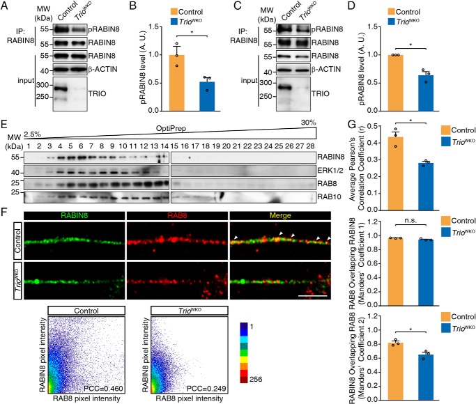
Figure 6.
TRIO promotes RABIN8 phosphorylation and regulates RABIN8 activity. A, RABIN8 phosphorylation analysis in cerebella tissues. B, quantification of the relative level of phosphorylated RABIN8 normalized to the pelleted RABIN8 in the blots shown in A. The error bars indicate S.E. (Student's t test). *, p < 0.05; n = 3. C, RABIN8 phosphorylation analysis in CGNs. D, quantification of RABIN8 phosphorylation in C. The error bars indicate S.E. (Student's t test). *, p < 0.05; n = 3. E, P10 mouse cerebellum was homogenized, and the postnuclear supernatants were subjected to 2.5–30% OptiPrep density gradient for subcellular fractionation. Fractions were subjected to Western blotting with RABIN8, RAB8, RAB10, and ERK1/2 antibodies. F, CGNs were cultured for 2 DIV and subjected to immunofluorescence using RABIN8 and RAB8 antibodies. The scale bar represents 5 μm. Scatter plots and the PCC of the fluorescence intensities of red and green channels were also shown. G, quantification of RABIN8 and RAB8 co-localization in neurites of CGNs as shown in F. Pearson's correlation coefficient and Manders' coefficient M1 and M2 were analyzed. The error bars indicate S.E. (Student's t test). *, p < 0.05. Each group comprised three mice (n = 3) and 18 neurons/group. A.U., arbitrary units; n.s., not significant; IP, immunoprecipitation; MW, molecular weight.

