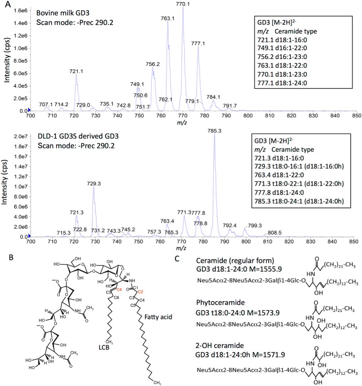
Figure 2.
Structure analysis of bovine milk- and DLD-1 GD3S-derived ganglioside GD3. A, identification of ganglioside GD3 structure by MS. Precursor ion scan of m/z 290 (the sialic acid–derived product ion in a negative ion mode) shows several molecular species of ganglioside GD3. Upper panel, mass spectrum of bovine milk–derived GD3. Lower panel, DLD-1 GD3-derived GD3. Carbon numbers of LCB were identified by MS/MS analysis (data not shown). B, long chain base C4 (red) is hydroxylated by DES2 (phytoceramide). Fatty acid C2 (red) is hydroxylated by FA2H (2-OH ceramide). C, structures of ceramide (regular form), phytoceramide, and 2-OH ceramide of GD3 are presented.

