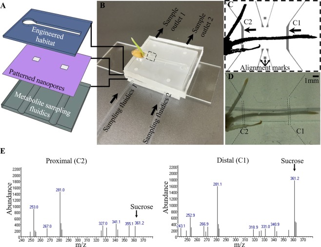
Figure 1.
Microfluidic device for simultaneous imaging of live root development and for metabolite sampling. (A) Schematic showing assembly of the three layers used to produce a culture system for a growing wheat root, infusion-patterned nanoporous membrane for sampling, and sample collection for analysis of metabolites. (B) Photograph of a germinated wheat seed growing in the device. (C) The primary root is placed in the sample microenvironment layer. The underlying sampling channels (C1 and C2) allow fluid pumping through ports, enabling buffer exchange and sampling of metabolites at two individually accessible locations. (D) Brightfield image showing the plant root in the primary microchannel after 6 h of growth in the device. The underlying metabolite sampling channels, highlighted by dashed lines, are obscured by the sandwiched nanoporous membrane. (E) Mass spectra derived from extracted-ion chromatogram (XIC) of sucrose indicating differential levels of sucrose in samples collected proximal and distal to the seed. The trimethylsilyl derivative of sucrose elutes at 15.09 min under the given separation conditions. The sucrose chromatographic peaks were determined by aligning with the given XIC for a sucrose standard. The Student t-test indicates that sucrose produced at the C1 and C2 locations are significantly different at P < 0.05 (95% confidence interval).

