Figure 2.
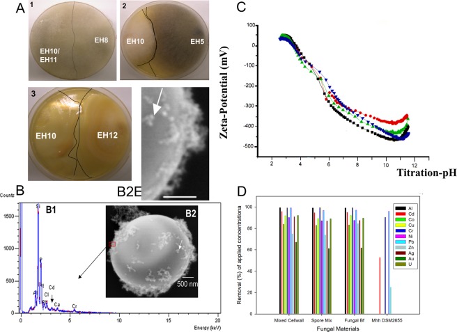
Inhibition/toxicity tests of M. hiemalis strains, detection of chitin, metal removal capacity and surface potential. (A1-3) Inhibition/toxicity tests of aquatic fungal strains, A1. Non-existence of demarcation lines between mycelial fronts showing absence of antagonistic inhibitory reactions among EH8, EH10 and EH11 when they were challenged against each other or grown together in the same plate, A2. Demarcation lines and discolouration indicating antagonistic reactions between EH5 and EH10, and A3. Demarcation lines and oily droplet formations illustrating antagonistic reactions between EH10 and EH12. (B-D) Relationship between metal binding and zeta-potential of the sporangiospore’s cell surface. (B) EDX detection of metals bound to the surfaces of sporangiospores, B1. EDX-detection of Al, Pb, Cd, Cr and P at a spot (red rectangle) on the outer surface of the sporangiospores (B1), B2 and B2E (enlarged). Formation of ca. 50–100 nanometer-sized particles (nanospheres; see white arrows) at the outer cell surfaces of sporangiospores following 48 h incubation in metal salt solutions (pH ∼7). (C) Zeta-potential of aquatic M. hiemalis sporangiospores after germination (1–3 cell stages) depending on nutrient conditions of incubation medium (red circle, C-limited medium; green triangle, C- and N-enriched medium; downward blue triangle, N-limited medium; black square, groundwater control) after 48 h incubation at approx. 30 °C. (D) Removal of metals by dead insoluble cell walls, live spore mix and live microbiomes (Fungal Bf) of strains EH8, EH10 and EH11 in comparison to the control terrestrial fungus DSM 2655. Horizontal bar in B2 and 2B2E indicates scale of 500 nm.

