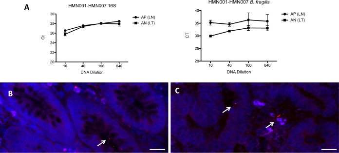
FIG 4.
Validation of the presence of Bacteroides fragilis in colonic tissues. qPCR amplification of microdissected DNA is shown. (A) Amplification of microdissected samples using 16S rRNA genes or B. fragilis primers. AP and AN represent the LCM identification of samples from the nontumoral (LN) and tumoral (LT) MAM regions. (B and C) Images are representative of FISH analyses with a B. fragilis-specific probe linked to Alexa 555 of a noncancerous tissue (B) or the paired tumoral colonic tissue (C) of the same patient. Nuclei are counterstained in blue with DAPI. Scale bars: 20 μm.

