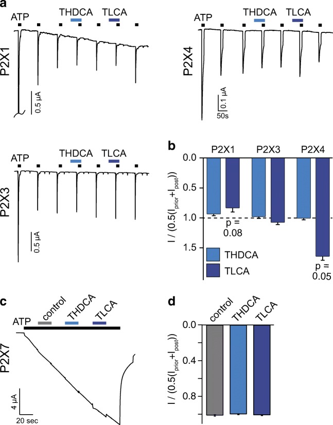
Fig. 3.
Only TLCA mildly modulated rP2X1 and rP2X4 currents. a Representative current traces of P2X1, P2X3, and P2X4 expressed in Xenopus laevis oocytes repetitively activated by ATP (P2X1, P2X3 0.3 μM; P2X4 3 μM). THDCA (20 μM) or TLCA (200 μM) (black bars) were pre- and co-applied as indicated by the blue bars. b Mean current amplitudes in the presence of the indicated bile acid relative to the average of the previous and the subsequent ATP-activated currents, n = 8–13. c Representative current trace of P2X7 expressed in Xenopus laevis oocytes activated by ATP (1.7 mM). Pre- and co-application of THDCA (20 μM) or TLCA (200 μM) (blue bars) or control (gray bar) did not affect current amplitude. d Mean current amplitudes in the presence of the indicated bile acid relative to the average of the current amplitude before and after bile acid coapplication. n = 6–9; error bars represent S.E.M.

