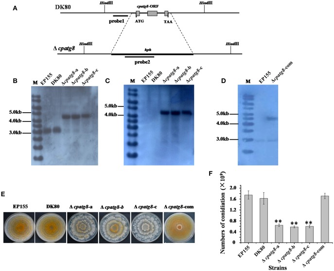
Figure 4.
Phenotypes and Southern analysis of cpatg8 knockout mutants. (A) Diagram of cpatg8 gene replacement strategy; probe fragment on the left arm was used in the Southern blot analysis to distinguish the fragment size of the wild-type strain and cpatg8 null mutants; Southern analysis of the cpatg8 null mutants (B,C) and complementary strain (D). Fungal total DNAs were digested with Hind III and separated on a 0.8% agarose gel by electrophoresis, and blotted using probe 1 (B) and probe 2 (C), respectively. Fragment sizes are indicated in the figure margins. (E) Mutant colony morphologies on PDA plates. Fungal strains were cultured on the laboratory bench top condition at 24°C, and the photograph was taken on day 14 postinoculation. (F) Sporulation level of cpatg8 knockout mutants. Spores were counted on day 14. Values are means ± S.E.M of three independent experiments. ** indicates P < 0.01, determined by Student's t-test.

