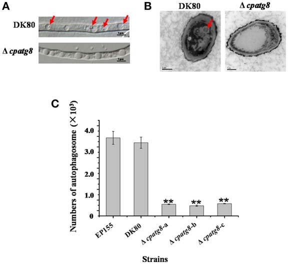
Figure 5.

Electron micrographs of the hyphae of strain DK80 and Δcpatg8 mutant. (A) Autophagy in the aerial hyphae of C. parasitica. Autophagic bodies in the vacuoles of the aerial hyphae of the strain DK80 and Δcpatg8 mutant grown on plates of PDA were examined using differential interference microscopy. (B) Autophagy was blocked in Δcpatg8 mutant. Vacuoles in the hyphae of the parental strain DK80 and Δcpatg8 mutant were observed using an electron microscope after being cultured in EP liquid media in the presence of 2 mM PMSF for 4 h (bar, 0.5 μm). (C) The quantification of the number of autophagic bodies. Arrow indicates autophagic body (bar, 5 μm). Values are means ± S.E.M of three independent experiments. ** indicates P < 0.01, determined by Student's t-test.
