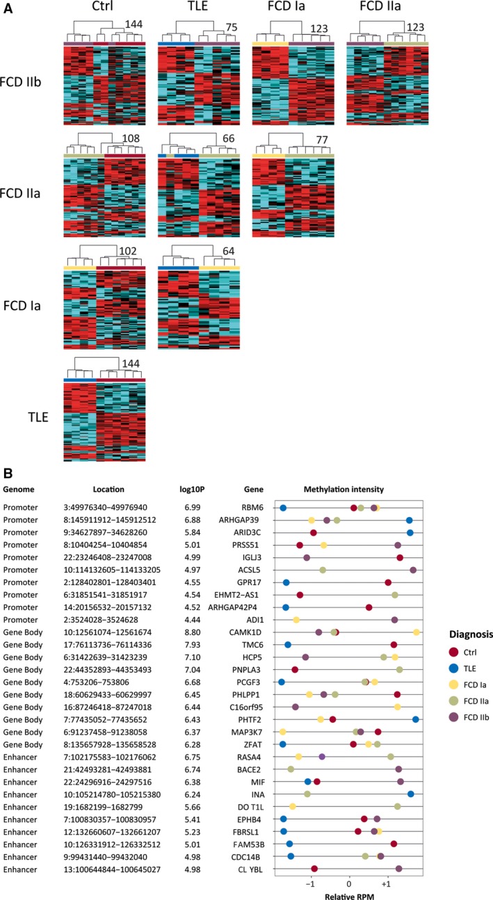
Figure 3.

A, Differentially methylated regions (DMRs) distinguished focal cortical dysplasia (FCD) Ia, FCD IIa, FCD IIb, temporal lobe epilepsy (TLE), and nonepilepsy control from each other in differential cluster analyses. Heatmaps of DMRs (P < 1e‐4) with low confounding variable influence (β < 0.5) are shown. The numbers of DMRs for each comparison are displayed next to each heatmap. B, Examples of differentially methylated genes (DMGs), which were identified through DMRs that are located at a gene's promoter, gene body, or enhancer. Genome, annotated part of the genome; Location, position of DMG on the hg19 human reference genome; log10P, the highest log10 P value for the DMG; Gene, annotated gene name; methylation intensity, scaled sequence abundance, showing comparisons with cutoff of P < 1e‐4; RPM, reads per million
