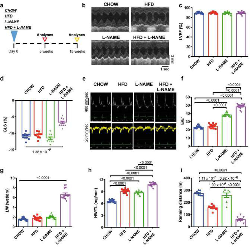
Figure 1. Fifteen weeks of HFD+L-NAME dietary regimen in mice recapitulates key alterations of clinical HFpEF.
a, Experimental design. C57BL/6N mice were maintained on different dietary regimens (filled triangle) and followed up to fifteen weeks (empty triangles). b, Representative left ventricular (LV) M-mode echocardiographic tracings. Images are representative of fifteen independent mice. c, Percent LV ejection fraction (LVEF%) (n=15 mice per group). d, LV global longitudinal strain (GLS) (n=10 mice per group). e, Representative pulsed-wave Doppler (top) and tissue Doppler (bottom) tracings. Images are representative of fifteen independent mice. f, Ratio between mitral E wave and E’ wave (E/E’) (n=15 mice per group). g, Ratio between wet and dry lung weight (LW) (n=15 mice per group). h, Ratio between heart weight and tibia length (HW/TL) (n=15 mice per group). i, Running distance during exercise exhaustion test (n=8 mice per group). Results are presented as mean±S.E.M. c, d, f-i One-way ANOVA followed by Sidak’s multiple comparisons test. Numbers above square brackets show significant P values.

