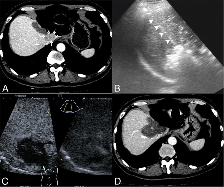
Fig. 2.

Images of a patient with recurrent hepatocellular carcinoma (HCC) in the caudate lobe treated by percutaneous radiofrequency ablation (RFA). a transverse contrast-enhanced computer tomography (CT) before treatment shows the tumor in the caudate lobe (arrow); b tumor appears completely hyperechoic after the beginning of RFA, and the electrode (arrowhead) is clearly showed on the ultrasound image; c CEUS image obtained 1 month after RFA shows the tumor completely ablated; d contrast-enhanced CT image obtained 1 month after RFA shows the tumor completely ablated
